
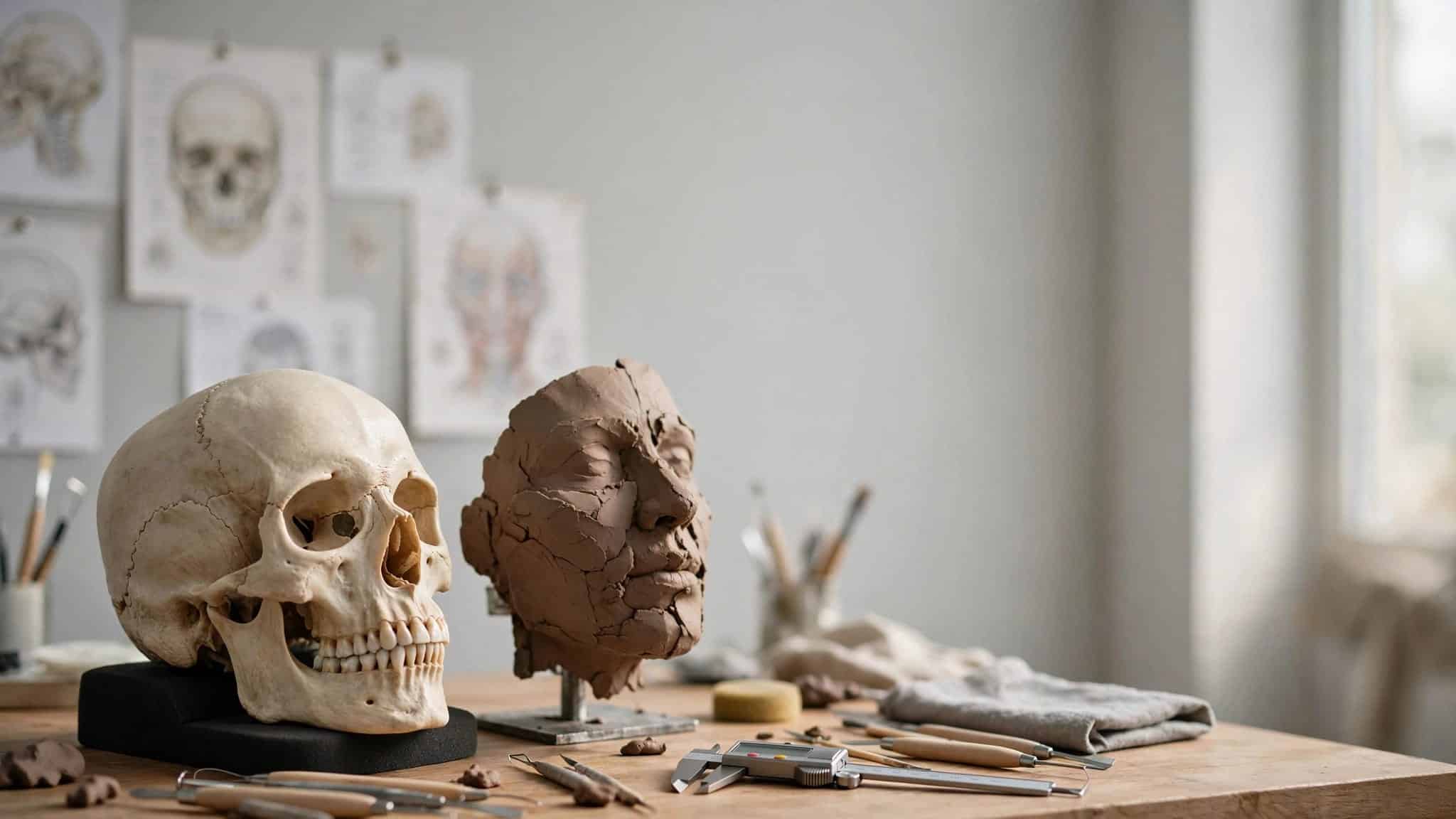
A skull can guide the rebuilding of a face, but it can’t preserve every detail that made that face familiar. Forensic facial reconstruction combines bone measurements, soft-tissue data and anatomical modeling to create an approximation of someone’s appearance.
For an ancient person, that can make the past feel surprisingly personal. For an unidentified person, it can encourage recognition and help investigators find a name. The process begins with understanding what the cranium can reveal, and what it leaves unanswered.
This post may contain affiliate links. If you make a purchase through these links, I may earn a small commission at no extra cost to you.
What forensic facial reconstruction actually tells us
Forensic facial reconstruction, also called facial approximation, recreates a possible face using skeletal remains. It relies on the relationship between bone, attached muscles, and the soft tissues beneath the skin.
The cranium constrains the face’s proportions. Cheekbones influence facial width, the jaw shapes the lower face, and the nasal opening provides clues about the nose. Those clues don’t specify one complete appearance.
This kind of forensic reconstruction can generate public recognition when other approaches haven’t produced an identity. It isn’t cosmetic surgery and doesn’t involve surgical tissue flaps. Research on facial approximation and identification distinguishes that role from positive identification.
That distinction matters when you see a finished reconstruction. A plausible likeness can help an investigation, but its lifelike appearance doesn’t make every detail equally well supported.
Preparing the skull without losing evidence
Before anyone adds a nose or a pair of cheeks, the remains need careful examination. Otherwise, a convincing face could be built on a mistaken starting point.
Establishing the person’s biological profile
In anthropological studies, forensic anthropologists use a morphological assessment to estimate age at death, biological sex and other characteristics. The wider skeleton can also help estimate stature. Ancestry assessment may contribute, although older literature’s term “race” oversimplifies human variation.
This biological profile narrows the possibilities; it doesn’t usually identify someone by itself. When the profile and conventional identification comparisons don’t resolve the case, facial approximation can provide another route toward recognition.
Preserving the original evidence
Ancient crania may be fragile, distorted or incomplete. Practitioners document those problems before deciding how missing areas should be restored. Physical work can use a replica, while CT scanning or surface scanning can produce a digital model.
Burial context matters too. As with dating ancient fossils, the surrounding layers help researchers interpret the remains. Rebuilding an appearance and establishing when someone lived are separate tasks, each requiring evidence.
Building soft tissue over bone
The reconstruction begins to look recognizable when tissue depth and anatomy come together. They set boundaries for facial features rather than provide a ready-made portrait.

Placing the tissue-depth markers
Practitioners position small pegs at standardized anatomical landmarks. Each peg estimates tissue thickness between the bone and skin’s surface. Digital reconstructions use virtual markers for the same purpose.
The relevant tissue thickness datasets are often grouped by age, sex and ancestry. They describe averages, so choosing a dataset doesn’t reveal an individual’s exact tissue depth.
The Office of Justice Programs’ survey of tissue-depth landmarks highlights the importance of standardizing these reference points. Measurements are more useful when practitioners agree about where they belong.
Adding the underlying anatomy
Depending on the method, the practitioner models facial muscles and other tissues around the skull. Muscle attachment sites help guide placement, while the markers constrain the outer surface.
Population coverage remains uneven. There isn’t a comprehensive tissue-thickness compendium representing every population worldwide, and group averages can’t capture every difference within a group.
That leaves room for judgment, particularly around body fat and age-related changes. The responsible approach is to document those choices rather than let them disappear beneath the finished skin.
Three ways to reconstruct a face
The main approaches differ in materials and workflow. Each depends on anatomical knowledge and decisions about uncertain features.
Two-dimensional drawings and composites
A practitioner can draw a face over a skull photograph or X-ray, or create a digital composite. The image needs an appropriate scale and orientation so the facial outline follows the underlying cranium.
This approach produces a flat representation rather than a head that can be viewed from different angles. Its usefulness depends on the quality of the source image and the reconstruction itself.
Manual three-dimensional sculpting
Manual reconstruction usually involves clay or plasticine applied to a cranium replica. The American approach traditionally emphasizes tissue-depth measurements, while the Russian anatomical approach emphasizes muscle reconstruction.
The Manchester method combines anatomical, muscle-by-muscle modeling with standard tissue-depth measurements. This hybrid gives the practitioner both an internal anatomical framework and guides for the skin’s surface.
The visible result may resemble portrait sculpture, but these artistic constructions are constrained by evidence rather than a sitter.
Computerized three-dimensional modeling
Virtual reconstruction builds tissues around a scanned cranium through virtual modeling. Tools can include modeling software such as FaceGen and haptic-feedback devices, which provide resistance as users manipulate virtual material.
A study of computerized facial approximation illustrates how researchers develop digital approaches. Virtual modeling lets researchers revise models without repeatedly handling fragile remains.
However, software doesn’t remove interpretation. A polished digital face can still contain uncertain choices about its nose, mouth or surface details.

Where the skull stops giving answers
Some features are constrained more closely than others. The nasal bones and opening offer clues about the nose, while the teeth and jaw help guide the mouth. Neither provides a complete record of the nose tip or lip shape.
Ears are particularly difficult because much of their visible form is cartilage. Hair, wrinkles, and expression also require interpretation or separate evidence. Eye color can’t be read from the cranium, although genetic evidence may sometimes help.
Hair and ears are facial features that strongly affect recognition. A hairstyle can make two versions of the same modeled face feel like different people.
A realistic finish can hide uncertainty: the most eye-catching details may be the least securely inferred from bone.
Cultural expectations create another risk. Ideas about beauty differ, so a universal, pleasing face template isn’t a sound substitute for individual anatomy. Reconstructions need to follow the person’s remains and facial structure, even when the result doesn’t fit a familiar artistic ideal. Balancing anatomical evidence with interpretation is the art of the science.
What historical faces can show us
Historical reconstructions are compelling because they put a human face beside a familiar name. However, knowing the name doesn’t automatically settle the appearance.
Dante Alighieri has been the subject of facial reconstruction work. The Dante Alighieri reconstruction shows why it’s important to distinguish virtual analysis of the cranium from the method used to produce a final face. A project can combine digital preparation with physical modeling, so calling the entire process “virtual” may leave out part of the work.
Research on Ferrante Gonzaga’s facial reconstruction used virtual anthropology to study the remains. For Ferrante Gonzaga, the digital approach let researchers examine anatomy without invasive handling and protect the original material.
For viewers, the helpful question is which details came from the cranium, which came from other evidence, and which required interpretation. That makes a historical face more informative without making it less interesting.
How a reconstructed face helps an investigation
In a cold case, an approximation may prompt someone to recognize a relative, friend or missing person. That response can generate a lead, but it isn’t forensic identification. Investigators pursue it through independent comparisons.
Mass disasters require the same separation between recognition and identification. INTERPOL’s disaster victim identification guidance emphasizes fingerprints, dental evidence and DNA. Facial reconstruction may help generate leads in unresolved cases, but it doesn’t replace those comparisons.
It’s also different from a police suspect composite. A composite relies on a witness’s facial recall, while reconstruction begins with skeletal evidence. Facial recall can be affected by memory contamination, sometimes called contagion, which is another reason to keep these practices distinct.
For families, an image may carry emotional weight long before an identification is established. Clear explanations of what the face can and can’t demonstrate help prevent a hopeful resemblance from becoming an unsupported conclusion.
Key takeaways
A cranium provides an anatomical framework, while tissue-depth measurements help estimate the face’s outer surface. Manual and digital methods use that evidence differently, but neither recovers every personal detail.
The strongest reconstruction makes its uncertainties visible. It separates features supported by bone from those informed by other evidence or artistic judgment. In an investigation, its practical purpose is to encourage recognition that can lead to further identification work.
Questions about rebuilding ancient faces
What happens if part of the cranium is missing?
Missing areas may be estimated using preserved anatomy, including the opposite side when appropriate. Those restorations introduce additional assumptions, which should remain documented.
The location of the damage matters. A missing section of the facial skeleton can affect the reconstruction differently from damage elsewhere. A complete-looking digital cranium may therefore contain both measured surfaces and estimated ones, even when the difference isn’t obvious in the finished image.
How can researchers assess whether a reconstruction worked?
Researchers can compare reconstructions with known faces or test whether viewers recognize the intended person. Surface measurements and recognition tasks assess different aspects of performance, so each gives a different view of the reconstruction’s degree of accuracy.
Research priorities include a consistent scale of accuracy for describing success, broader tissue-thickness datasets and software that connects those data to virtual models. These improvements help make methods easier to evaluate rather than simply more visually persuasive.
Rebuilding a face while respecting the unknown
Reconstructing an ancient face requires anatomical knowledge, measurements and careful interpretation. The cranium provides meaningful constraints, but some personal details remain beyond its reach.
A responsible likeness makes that balance clear. It helps us picture an individual while preserving the difference between what the evidence supports and what someone had to imagine.


