The phase contrast microscope is an optical microscope that is used to observe transparent samples, such as living cells, without staining or fixing them. It enhances the contrast of the sample by converting the phase shift caused by differences in refractive index of the sample into amplitude changes. In this article, we will discuss how to use a phase contrast microscope to observe transparent samples.
Principle of Phase Contrast Microscopy
The principle of phase contrast microscopy is based on the fact that light waves passing through a transparent sample are delayed or phase-shifted by the different refractive indices of the sample components. This phase shift cannot be detected by the human eye or a standard microscope, as the waves have the same brightness and color. However, by introducing a phase plate into the microscope, the phase shift can be converted into amplitude differences, which are visible as contrast.
Sample Preparation
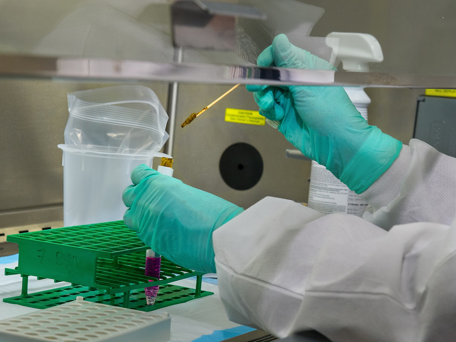
Sample preparation for phase contrast microscopy is relatively simple, as the technique allows for observation of live, unstained samples. The sample is placed on a glass slide and covered with a coverslip to prevent it from drying out. For best results, the sample should be as thin as possible, preferably one cell layer thick. If the sample is too thick, the light waves passing through it will be scattered and the image will be blurred.
Image Acquisition
To observe a sample using a phase contrast microscope, the microscope is set up with the appropriate phase plate and objective lens. The phase plate introduces a phase shift of 0.25 to 0.5 wavelengths of light, which causes the phase shift in the sample to be converted into amplitude differences. The objective lens focuses the light onto the sample and collects the scattered light. The image is then magnified and focused onto the eyepiece or camera.
Image Processing
Image processing is not usually required for phase contrast microscopy, as the technique produces high contrast images without the need for staining or other modifications. However, the images can be adjusted for brightness and contrast, and annotations can be added using image processing software.
Applications
Phase contrast microscopy is used in a variety of fields, including biology, medicine, and materials science. In biology, it is used to study the structure and behavior of living cells, including the movement of organelles and the formation of cell structures. In medicine, it is used to diagnose diseases, such as malaria, and to study the behavior of pathogens, such as bacteria and viruses. In materials science, it is used to study the structure and properties of transparent materials, such as polymers and glasses.
Conclusion
Phase contrast microscopy is a powerful technique that allows for the observation of transparent samples, such as living cells, without staining or fixing them. It enhances the contrast of the sample by converting the phase shift caused by differences in refractive index of the sample into amplitude changes. Sample preparation is relatively simple, and image acquisition produces high contrast images without the need for staining or other modifications. Phase contrast microscopy has many applications in biology, medicine, and materials science, making it an important tool for scientific research and discovery.


