The pituitary gland, also known as the hypophysis, is a small, pea-sized gland located at the base of the brain. It is often referred to as the “master gland” because it controls the function of other endocrine glands in the body. Despite its small size, the pituitary gland plays a critical role in maintaining a wide range of bodily functions, including growth and development, metabolism, and reproductive function.
Identifying the Location of the Pituitary Gland in the Brain
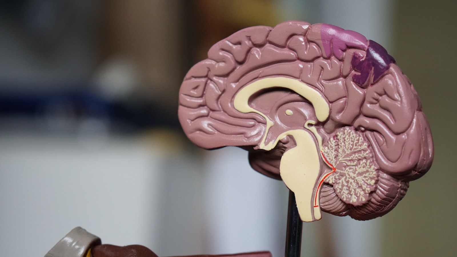
Anatomy of the Pituitary Gland
The pituitary gland is located in a small depression in the sphenoid bone called the sella turcica. It is divided into two distinct lobes: the anterior pituitary gland, also known as the adenohypophysis, and the posterior pituitary gland, also known as the neurohypophysis.
The anterior pituitary gland is larger and more complex than the posterior pituitary gland. It is composed of glandular tissue and produces and releases six different hormones that regulate the function of other endocrine glands in the body. The hormones produced by the anterior pituitary gland include adrenocorticotropic hormone (ACTH), follicle-stimulating hormone (FSH), luteinizing hormone (LH), growth hormone (GH), prolactin, and thyroid-stimulating hormone (TSH).
The posterior pituitary gland, on the other hand, is made up of nervous tissue and stores and releases two hormones produced by the hypothalamus: oxytocin and vasopressin (also known as antidiuretic hormone or ADH). The posterior pituitary gland does not produce any hormones of its own.
Identifying the Pituitary Gland
The pituitary gland is located at the base of the brain, behind the eyes and below the hypothalamus. Because of its location deep within the brain, it cannot be directly visualized or palpated like other organs in the body. However, there are a few ways to identify the location of the pituitary gland:
Imaging techniques: Modern imaging techniques, such as magnetic resonance imaging (MRI) and computed tomography (CT) scans, can provide detailed images of the brain and its structures. These scans can be used to visualize the pituitary gland and its surrounding structures and identify any abnormalities or tumors.
Nasal endoscopy: Another way to visualize the pituitary gland is through nasal endoscopy. This procedure involves inserting a thin, flexible tube with a camera on the end through the nose and into the back of the throat. The camera can then be used to view the pituitary gland and surrounding structures.
Symptoms of pituitary disorders: Disorders of the pituitary gland can cause a variety of symptoms, depending on which hormones are affected. For example, a deficiency in growth hormone may cause slow growth and short stature, while excess prolactin production may cause breast milk production in non-pregnant women. Identifying these symptoms can lead to a diagnosis of a pituitary disorder and subsequent imaging or other diagnostic tests.
Conclusion
The pituitary gland is a small but critically important gland located at the base of the brain. While it cannot be directly visualized or palpated, modern imaging techniques and other methods can be used to identify its location and diagnose any disorders or abnormalities. Proper identification and diagnosis of pituitary disorders is important in order to properly treat and manage these conditions and maintain overall health and well-being.


